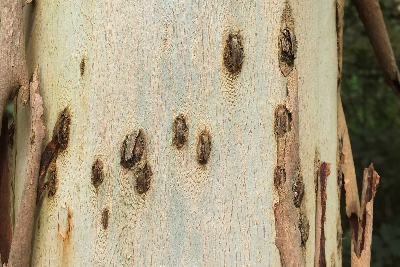
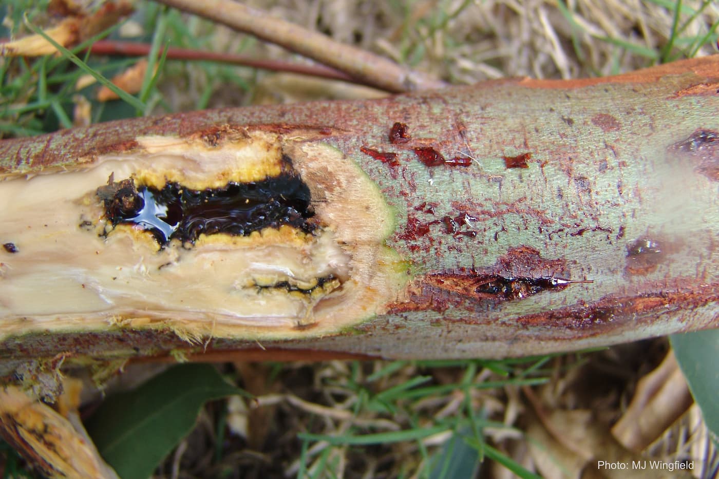
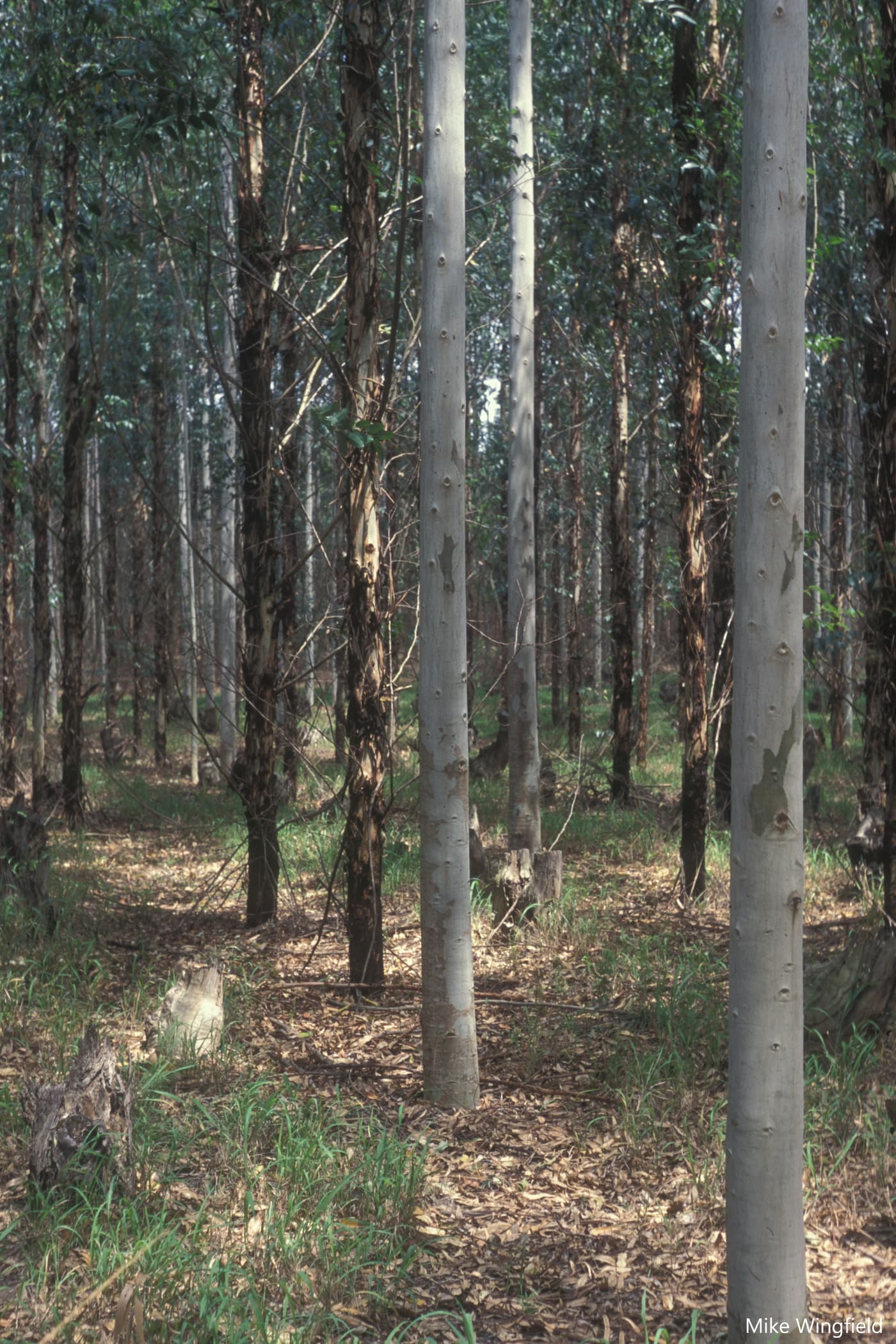
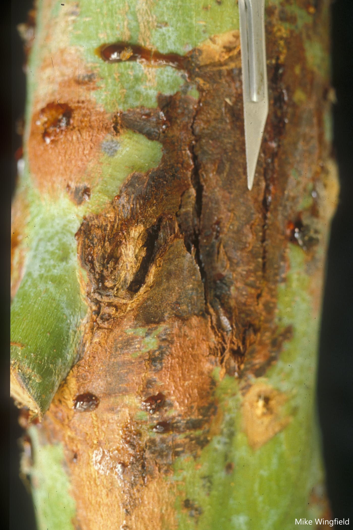
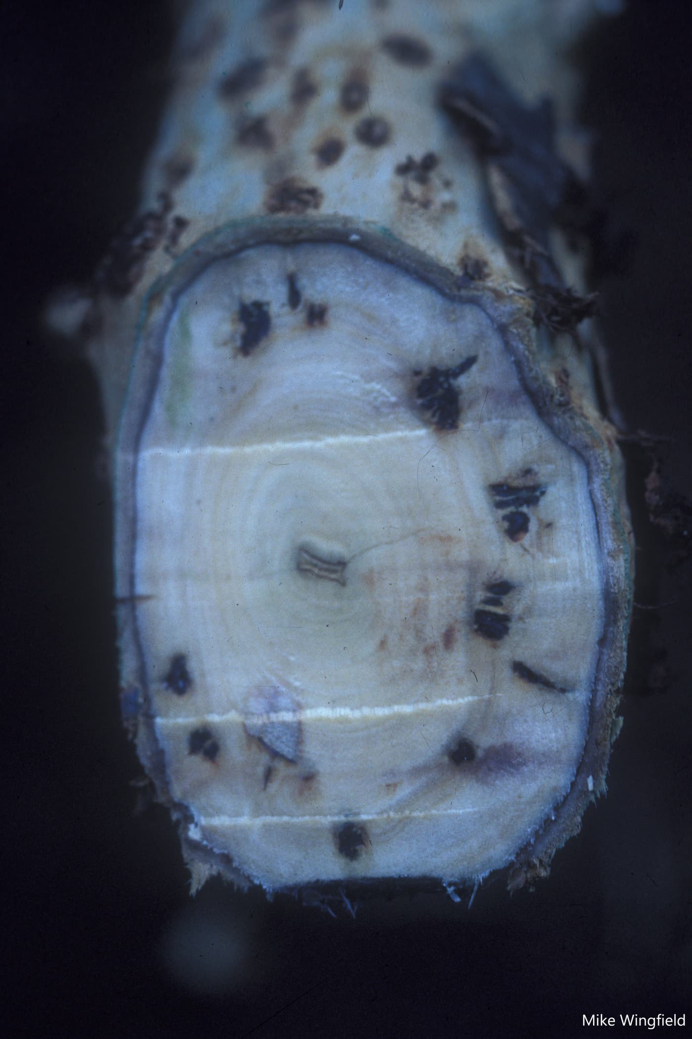
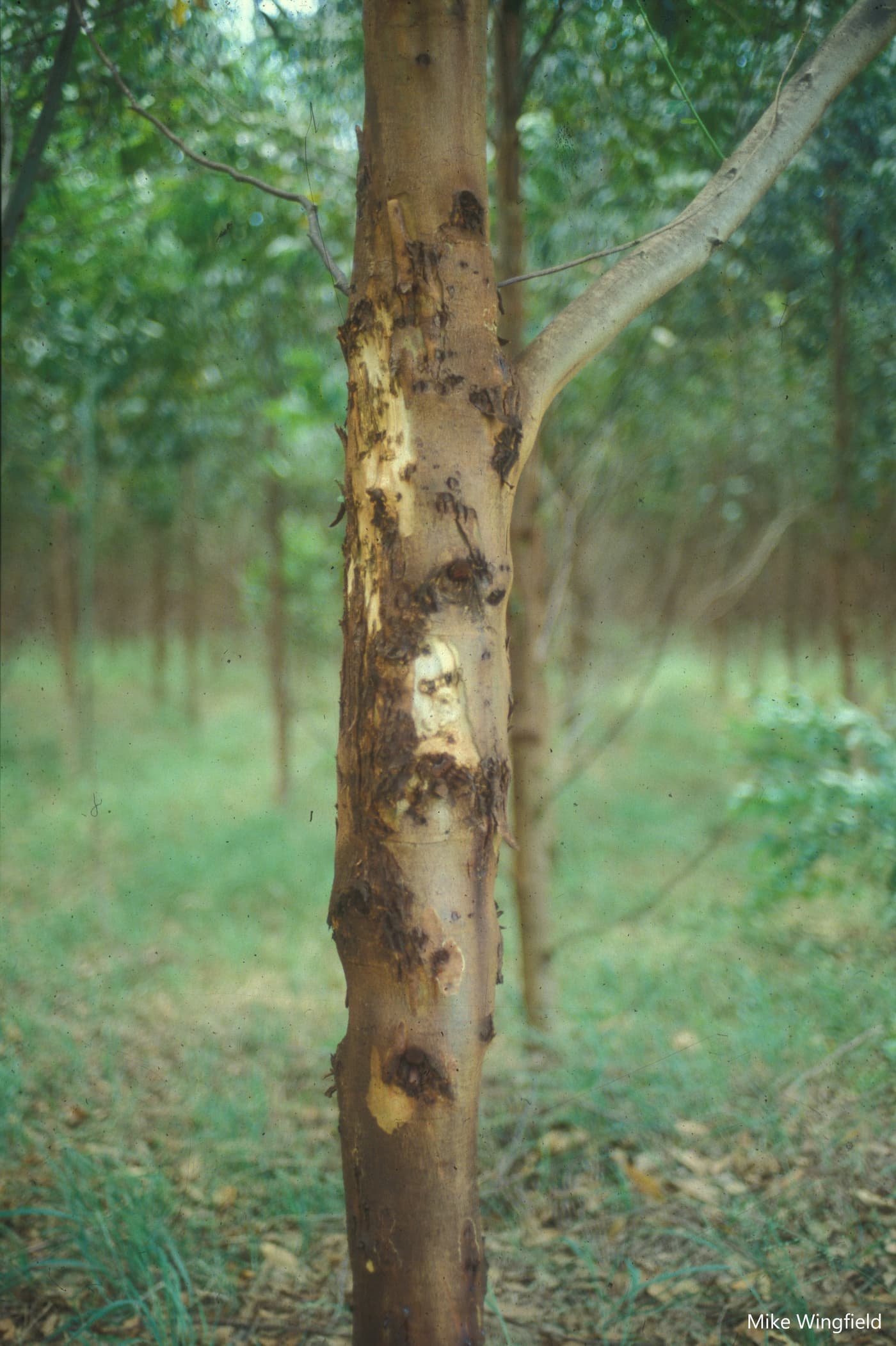
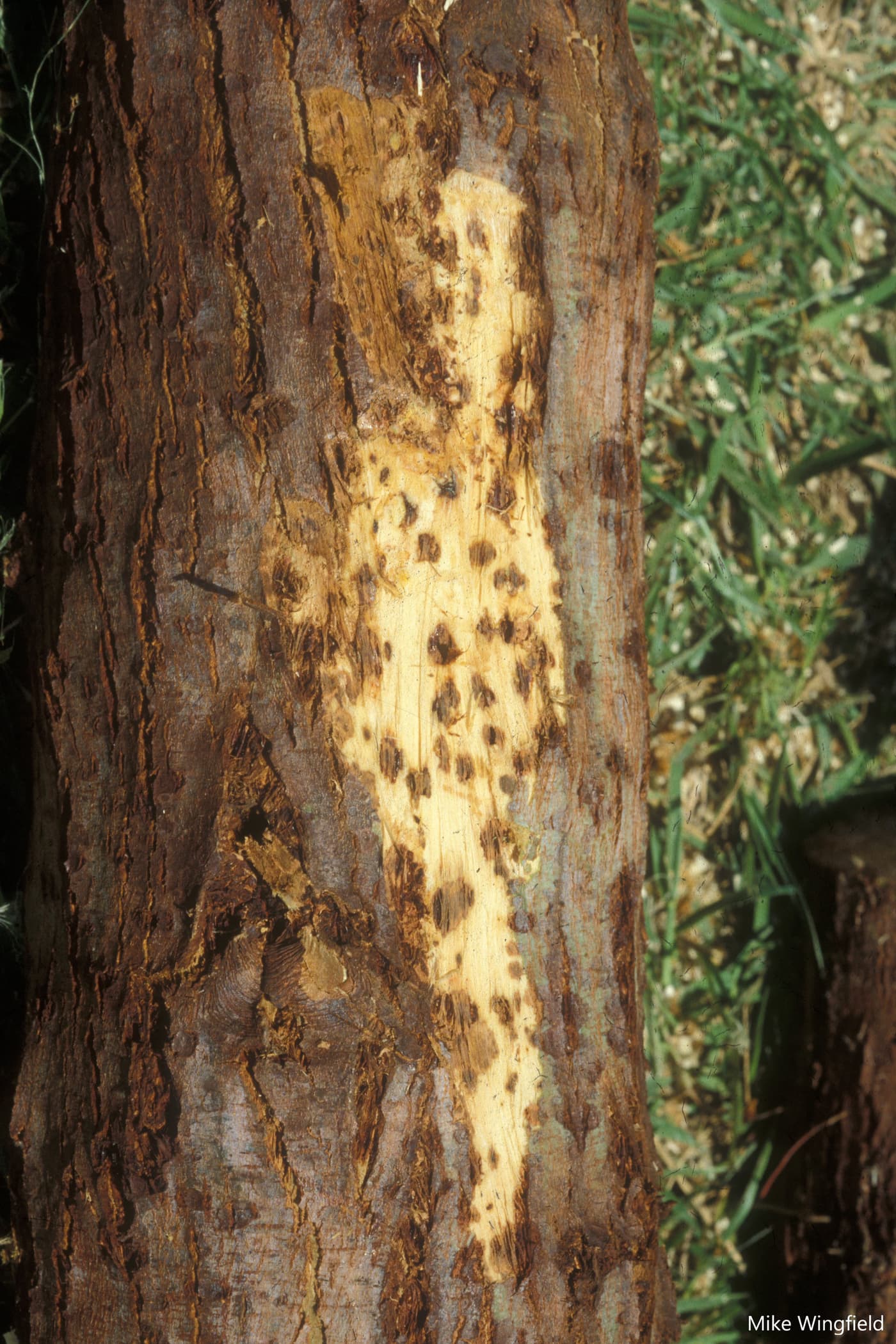
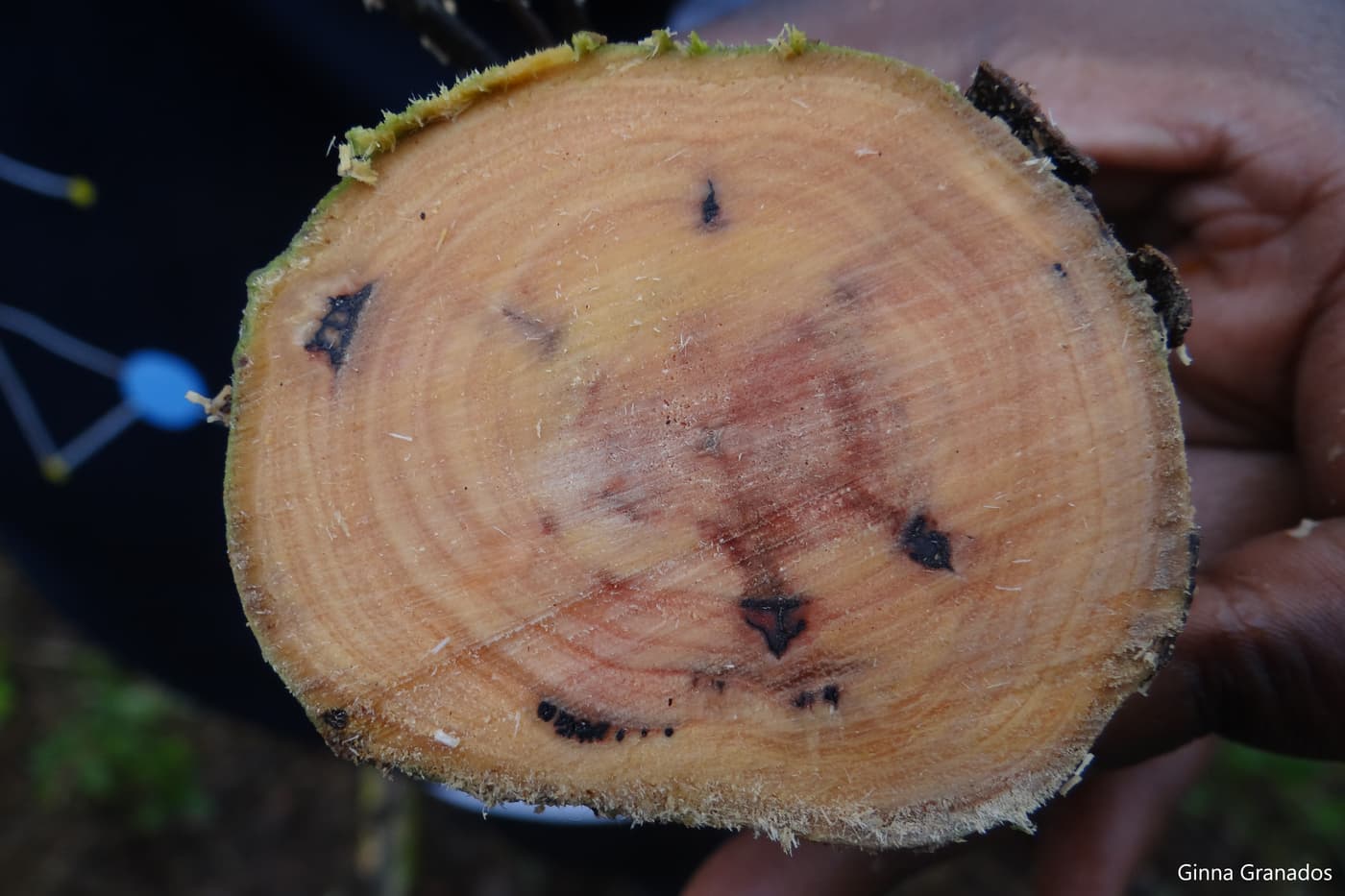
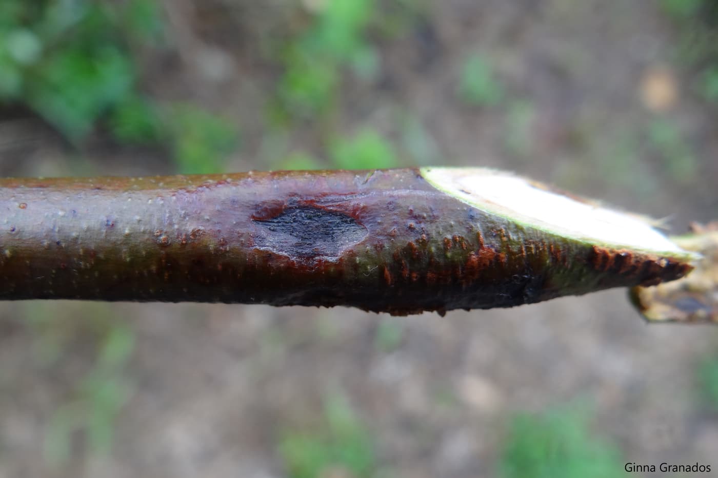
Teratosphaeria gauchensis and T. zuluensis cause indistinguishable disease symptoms (Aylward et al. 2019). Initial infections occur on young, green stem tissue and are first visible as small (2-5 mm) lesions on bark (Wingfield et al. 1996). Lesions become elliptical as their size increases, they penetrate the vascular cambium and eventually merge with neighbouring lesions to form cankers filled with gum, also known as kino pockets (Wingfield et al. 1996; Old et al. 2003). The bark covering these cankers often cracks vertically, creating a “cat-eye” appearance and causing the gum to exude (Cortinas et al. 2006). In the case of severe infections on susceptible clones, cankers girdle the stems, epicormic shoots develop and the tops of the trees die (Wingfield et al. 1996; Old et al. 2003).
Tree Protection Co-operative Programme
Teratosphaeria stem canker (previously Coniothyrium)
Download PDFTeratosphaeria gauchensis (M.-N. Cortinas, Crous & M.J. Wingf.) M.J. Wingf. & Crous and Teratosphaeria zuluensis (M.J. Wingf., Crous & T.A. Cout.) M.J. Wingf. & Crous
Symptoms
Biology
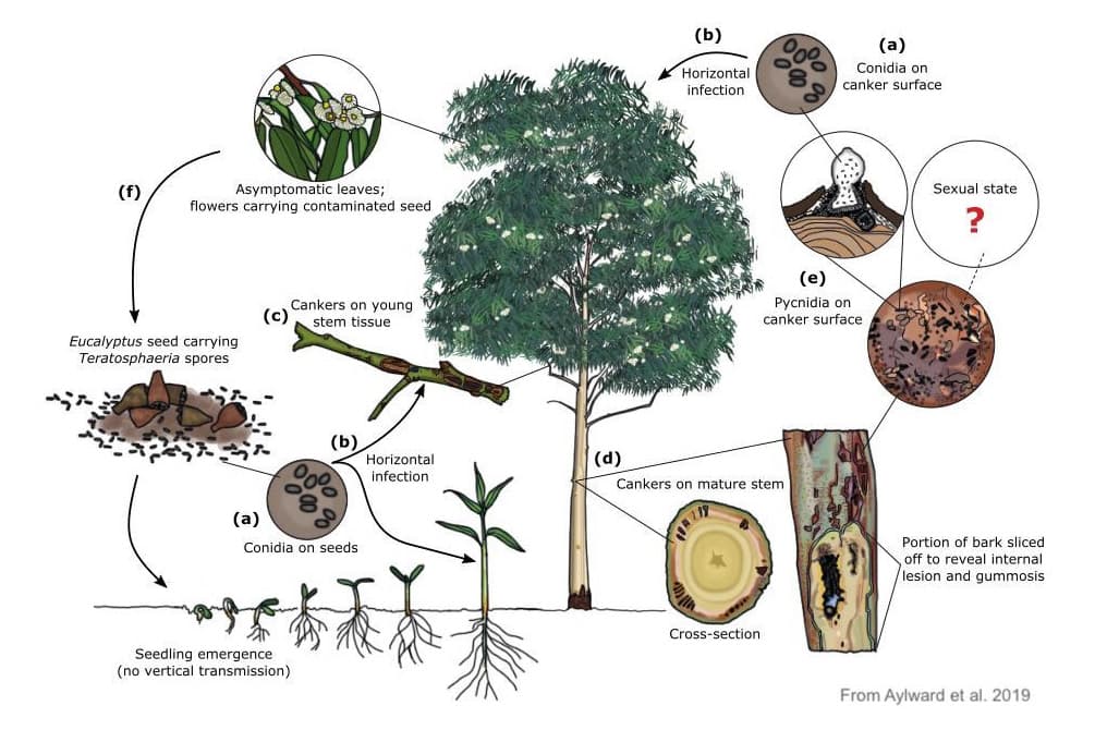
Little is known about the biology of T. gauchensis and T. zuluensis, but evidence gathered during the course of the past two decades provides a likely scenario of their lifecycle in Eucalyptus plantations (Aylward et al. 2019).
Both T. gauchensis and T. zuluensis are present in Eucalyptus leaves (Pérez et al. 2009; Marsberg et al. 2014) and may also occur in the seeds and seed capsules of infected trees (Jimu et al. 2016). Seedlings that come into contact with contaminated plant debris are infected horizontally after they begin to grow. Infection of the green tissues of Eucalyptus trees most probably occurs from conidia produced on cankers and other plant tissues carrying asymptomatic infections.
Analysis of the available whole genome sequences of these species (Wingfield et al. 2019) has revealed that both follow a heterothallic mating strategy. Isolates of T. gauchensis and T. zuluensis will, therefore, have either the MAT1-1 or MAT1-2 mating type (Aylward et al. 2020). There is no physical evidence of sexual reproduction for either species and recombination is unlikely to form an important part of their life cycles in diseased Eucalyptus plantations.
Gallery